SIGN UP TO RECEIVE
15% OFF
IN YOUR NEXT TOUR
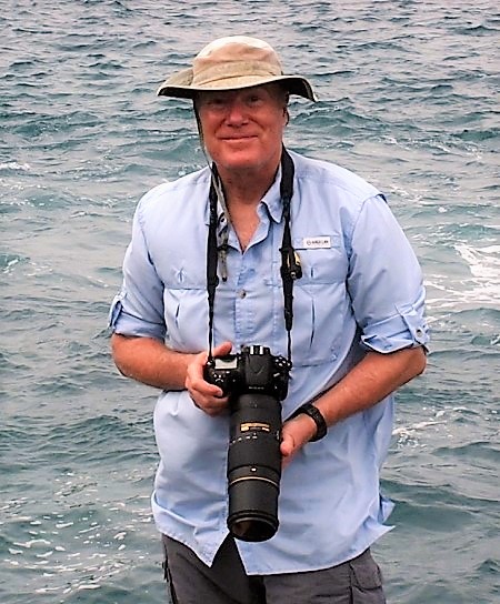
Jim Wetzel
Biologist, Naturalist, Photo Instructor

As a professor of biology at a liberal arts college his classes are often a mix of student across various disciplines – science, the humanities, and the visual arts. But a common theme throughout is developing one’s personal connection with the earth. He developed Environmental Photography as a course and workshop to help students develop their own photo essays of personal expression and environmental awareness.
His doctoral degree is in Zoology, specifically working on marine embryos, and He employs primarily light and electron microscopy for research. Through microscopy as a research tool, He developed an interest in the invisible side of nature – forms not seen by the unaided eye. This ‘invisible world’ has now become a regular theme within my Environmental Photography workshops, and one that students seem to enjoy at times even more than conventional imagery. Viewing the microscopic helps complete their appreciation of the interconnected web all of nature’s forms.
His current research involves three aspects: 1) developmental anatomy, 2) materials transfer in viviparous fishes, and 3) correlative microscopy using both fluorescent and electron microscopy and x-ray diffraction microanalysis. For reach of these He has a substantial background that is primarily based on years of research with undergraduate biology majors.
As regards developmental anatomy (1) – this is His primary interest, and served as the greater part of His graduate school training and the focus of His doctoral dissertation involving functional morphology of the seahorse brood pouch.
The seahorse brood (2) – pouch functions on a very basic level for sequestering ions in seawater and material such as iron that is transported from the paternal tissues to the developing embryo.
His research methods (3) – have always employed both light and electron microscopy – and with the growing interest in recent years in correlative microscopy studies, He combine both light and electron microscopy with x-ray diffraction analysis of materials stored in the parental seahorse tissues and along the embryonic gut in the embryo.
– Biocommunications Association award: Photomicrography “Image of Merit” (2018)
– Canadian Founders Natural Science award, 1st place (2010)
– Nikon Small World competition, “Image of Distinction” (2010)
– Biocommunication Association award: Photomicrography: “Image of Merit” (2010)
– Olympus Bioscapes Competition – honorable mention (2005)
– EIPBN Grand Prize winner – electron microscopy competition (2004)
– Microscopy Today journal: front cover electron micrograph image (2001)
– Polaroid Corporation International Scientific Photographic Competition – First Place (1994)
bca.org/publications/news/winter2019/dragons/html
